Browse products by medical specialities area or therapies
Product by Therapies
Product by Type
Product by Therapies
Product by Type
Arthroplasty
Product by Therapies
Product by Type
Sports Medicine
Product by Therapies
Knee Portfolio - ACL /PCL Reconstruction
Knee Portfolio - Meniscus Repair
Shoulder Portfolio - Bankart/Slap Repair
Shoulder Portfolio - Rotator Cuff Repair
Product by Type
Selecting a tibial implant is not simply a matter of choosing the newest design. It requires careful attention to fixation, alignment, polyethylene compatibility, and patient anatomy. In this guide, “Tibial implant rotational stability” is the central evaluation theme. We examine ten leading tibial implant options available in China, focusing on how their geometry may resist unwanted movement after knee replacement.
The review considers practical details that influence rotational control. These include keel or stem design, tray coverage, cortical support, porous surfaces, locking mechanisms, and instrument accuracy. Surgical technique remains critical. Even a well-designed component may perform poorly when sizing, rotation, or soft-tissue balancing is inaccurate. Experienced surgeons also assess bone quality, deformity, activity level, and revision risk before selecting an implant.
Evidence matters here. The comparison should draw from peer-reviewed studies, clinical follow-up, registry data, manufacturer documentation, and applicable NMPA requirements. Laboratory findings can be useful, but they do not always predict long-term patient outcomes. That distinction deserves attention.
No ranking is universally correct. Implant availability, surgeon familiarity, hospital protocols, and patient anatomy can change the final choice. Some designs may appear highly stable in testing yet offer limited independent clinical evidence. Others have strong clinical use but less transparent comparative data. This guide therefore presents a structured, cautious review rather than a definitive verdict. Readers should confirm current approvals, indications, and surgical instructions before clinical adoption.
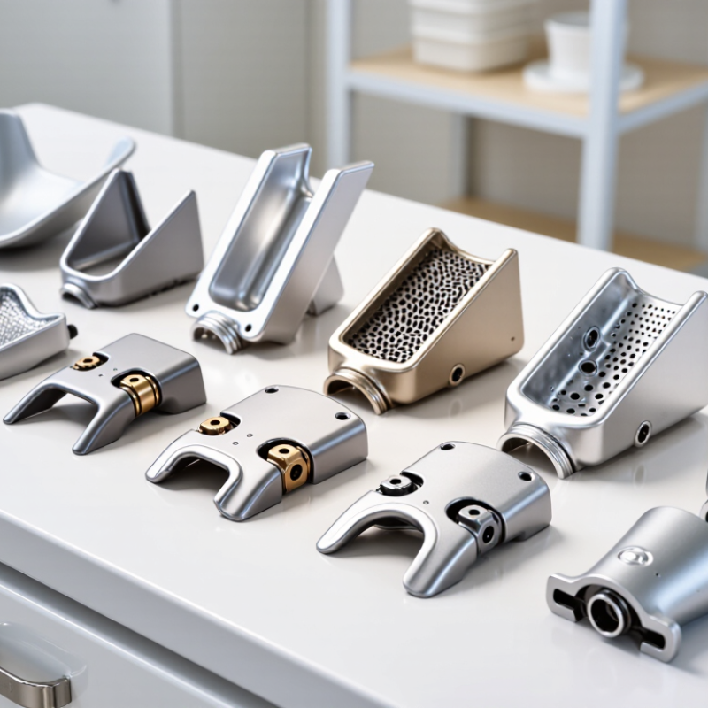
Rotational stability describes how well a tibial implant resists twisting after implantation. It is not the same as simple vertical fixation. Three measures matter: micromotion, torsional rigidity, and tibial fit. Micromotion is tiny movement at the bone-implant interface. Excessive movement may disturb bone ingrowth and contribute to pain or loosening. Laboratory thresholds can guide screening, but patient bone quality changes their meaning. Real knees are less predictable.
For a China-focused top-ten comparison, testing should combine mechanical data with clinical evidence. Torsional rigidity shows how strongly the tibial tray resists rotational loading during walking, stair climbing, and rising from a chair. A very rigid design is not automatically better. It may transfer stress unevenly if the bone is weak or the load path is poorly matched. I would examine cadaveric testing, follow-up duration, revision rates, and study transparency before accepting a ranking.
Tibial fit deserves equal attention. The component should cover supportive cortical bone without excessive overhang. Poor coverage can leave gaps; overhang may irritate surrounding soft tissue. Trial components, radiographs, and patient-specific anatomy help reveal these problems. Stem position also matters, especially when the metaphysis is irregular. My own first-pass assessments can overvalue impressive laboratory numbers. That is a useful weakness to acknowledge. A reliable review should compare micromotion under repeated loading, torsional behavior, and fit across varied tibial shapes, not just one ideal specimen.
The chart compares ten generic tibial implant design profiles using a normalized 0–100 evidence-informed index. Higher values indicate lower expected micromotion, greater torsional rigidity, or better tibial coverage and fit. These are design-level comparisons rather than manufacturer rankings or clinical product claims.
Micromotion below approximately 150 μm is commonly used as a benchmark associated with improved potential for bone ingrowth in cementless fixation. Actual rotational stability depends on bone quality, implant positioning, cortical support, surface coating, fixation method, and patient-specific anatomy. Clinical selection should be based on peer-reviewed evidence and regulatory documentation.
Reference concepts: RSA-based migration studies, cementless fixation literature, tibial component coverage studies, and biomechanical assessments of torsional rigidity.
Evaluating China’s top 10 tibial implants for rotational stability requires more than a brochure review. A credible process should combine ISO 14243 wear testing with radiostereometric analysis, or RSA. These methods answer different clinical questions.
ISO 14243 testing simulates repeated knee loading, flexion, and sliding motion. Test reports should show lubricant conditions, load profiles, cycle numbers, insert material, and measured wear. Mass loss alone is insufficient. Investigators should examine wear patterns, deformation, and rotational behavior under changing loads. A tibial component that performs well on a machine may still behave differently in a patient.
RSA adds the clinical dimension. Radiopaque markers can track tibial tray migration and rotation with high precision. Early rotational migration deserves attention, especially during the first two years after implantation. Stable motion matters. Researchers should report measurement error, follow-up intervals, fixation method, and patient activity levels. Without these details, comparisons become weak.
No test is perfect. ISO 14243 cannot reproduce every bone quality or surgical technique. RSA also depends on marker placement and image quality. I would avoid ranking implants from one dataset alone. A stronger selection combines standardized wear results, RSA stability data, manufacturing traceability, and independently reviewed clinical evidence. That approach is slower, but more defensible.
China Top 10 Tibial Implants for Rotational Stability?
Comparing ten Chinese tibial implant designs requires more than checking tray size or surface finish. Keel and peg geometry often controls rotational resistance, especially during early loading. A deep central keel can improve axial fixation, while a wider fin may resist tray spinning. Shorter keels may preserve bone, but they can provide less control in softer metaphyseal bone. Fit matters.
Among these ten designs, several use one central keel with broad anterior-posterior support. Others combine a central keel with two or more pegs. Tapered pegs can guide insertion and reduce local stress concentration. Cylindrical pegs may offer consistent fixation, yet their performance depends heavily on preparation accuracy. Stepped peg profiles may improve purchase in selected bone conditions, but they are not automatically superior. Small details matter.
From a clinical assessment perspective, rotational stability should be reviewed with radiographs, component sizing, bone quality, and operative technique. Cemented and cementless fixation also changes how keel and peg features perform. Public comparative evidence remains uneven across Chinese implant systems. That limitation deserves attention. A ranking is never absolute. Surgeons should examine tibial coverage, posterior slope, keel depth, extraction options, and documented clinical follow-up. One design may feel stable during trialing but reveal poor coverage after final positioning. This is where careful measurement, experienced judgment, and honest review become essential.
| Profile | Primary Tibial Fixation | Keel / Peg Configuration | Rotational-Stability Mechanism | Typical Design Rationale | Potential Advantages | Key Considerations | Overall Design Emphasis |
|---|---|---|---|---|---|---|---|
| Design Profile 01 | Central keel | One broad, longitudinal central keel with anterior–posterior orientation | Resistance to torsional movement through keel-bone contact and a broad baseplate footprint | Simple, conventional geometry intended for primary cemented fixation | Familiar instrumentation concept; relatively straightforward bone preparation | Rotational control depends strongly on keel width, cement mantle, cortical support, and bone quality | Balanced |
| Design Profile 02 | Twin keel | Two parallel keels separated by a central channel | Increased resistance to rotation through a wider transverse stabilizing geometry | Distributes torsional loads across two longitudinal fixation features | May improve resistance to baseplate rotation when adequate cancellous support is present | Requires accurate keel-slot preparation and sufficient tibial bone stock | High rotational focus |
| Design Profile 03 | Four-peg fixation | Four short pegs positioned around the central load-bearing area | Multiple point contacts limit rotational and rocking motion | Uses distributed fixation rather than one large central keel | Can provide broad peripheral fixation and a comparatively low central bone-preparation profile | Peg placement accuracy is important; small pegs may be less forgiving in weak bone | Distributed fixation |
| Design Profile 04 | Keel plus pegs | Central keel combined with two or more supplemental peripheral pegs | Combines longitudinal keel resistance with multi-point anti-rotation support | Designed to share torsional and vertical loads between central and peripheral features | Redundant fixation pattern may reduce dependence on one single fixation element | More extensive preparation can increase surgical complexity and bone-removal requirements | Enhanced stability |
| Design Profile 05 | Long stemmed keel | Extended central keel or short stem projecting farther into the proximal tibia | Longer bone engagement increases the lever arm opposing rotational displacement | Intended to improve load transfer beyond the immediate tibial cut surface | Potentially useful where additional axial and torsional guidance is required | Alignment, stem position, and compatibility with the patient’s tibial canal are critical | Deep fixation |
| Design Profile 06 | Short keel with peripheral pegs | Low-profile central keel supported by anterior, posterior, or posterolateral pegs | Peripheral peg spacing creates a wider rotational control envelope | Seeks to balance fixation with bone preservation around the central tibia | Lower central preparation may preserve more cancellous bone than a large keel | Peripheral bone quality and accurate rotational positioning are important | Bone-preserving balance |
| Design Profile 07 | Asymmetric keel | Keel geometry offset or shaped to follow the medial–lateral tibial anatomy | Improved cortical support and asymmetric bone engagement may reduce rotational micromotion | Designed to accommodate the non-circular shape of the proximal tibial resection | Can improve coverage and reduce unsupported plate overhang when sizing is appropriate | Incorrect rotation or sizing may compromise coverage and soft-tissue clearance | Anatomy-oriented |
| Design Profile 08 | Porous-coated keel | Central keel or stem with a porous or textured surface for biological fixation | Initial geometric stability is supplemented by long-term bone ingrowth where conditions permit | Intended primarily for cementless fixation protocols | Potential for durable biological fixation in patients with suitable bone quality | Early stability remains essential; ingrowth performance depends on bone biology, alignment, and micromotion control | Cementless fixation |
| Design Profile 09 | Three-post fixation | Three separated posts or pegs arranged in a triangular support pattern | Triangulated fixation resists rotation and rocking in multiple directions | Uses non-collinear fixation points to improve mechanical stability | Can provide directional stability without the preparation required for a long keel | Requires careful preparation of each post site and adequate metaphyseal bone support | Multi-directional control |
| Design Profile 10 | Conical stem with keel | Central keel transitioning into a tapered or conical short stem | Tapered stem engagement provides axial guidance and additional resistance to rotation | Combines surface fixation with metaphyseal canal engagement | May improve alignment control and load transfer in appropriately sized tibiae | Stem position, canal preparation, and avoidance of cortical impingement require attention | Alignment and fixation |
China’s top 10 tibial implants for rotational stability should be judged by fixation data, not catalogue language.
The key question is simple: does the tibial component remain below 150 μm of micromotion under functional loading? This threshold is widely used as a practical benchmark for bone ingrowth, but it is not a universal guarantee. Surface structure, press-fit geometry, bone quality, and loading direction can change the result. Small numbers matter.
In a serious comparison, I would examine RSA or validated optical tracking, test protocols, and follow-up duration. A bench result at 2,000 newtons cannot be compared directly with a six-week clinical measurement.
Rotational movement deserves separate attention. A component may show acceptable vertical migration yet rotate around the keel during stair climbing. Look for cyclic loading, varus-valgus challenges, and low-density bone models. The raw micromotion range is more informative than a single average.
Clinical evidence should report patient selection, revision rates, pain scores, and radiographic changes. Independent investigators add credibility, especially when methods and adverse events are disclosed. I would also question polished rankings. Some studies use small samples, inconsistent landmarks, or short observation periods. That weakens certainty. A practical shortlist should favor reproducible fixation below 150 μm, stable rotation, and transparent testing over impressive marketing claims.
China’s top 10 tibial implants should be ranked by evidence, not advertising. A practical score can assign 40% to alignment accuracy, 40% to revision rates, and 20% to follow-up quality. Designs with independent radiographic studies should outrank systems supported only by laboratory testing. Long-leg imaging matters. A two-degree varus error can change load distribution across the knee.
Registry data provide a useful benchmark. The Australian Orthopaedic Association National Joint Replacement Registry 2024 report places overall primary knee replacement revision at roughly 5–6% at ten years. The UK National Joint Registry’s 21st Annual Report also shows meaningful differences by patient age, fixation method, diagnosis, and implant size. These reports do not prove one tibial design is best in China. They show why revision outcomes need risk adjustment.
Alignment evidence remains mixed. Recent comparative studies report fewer mechanical-axis outliers with navigation or robotic assistance, yet improved accuracy has not consistently produced lower revision rates. That gap deserves attention. For a Chinese top-ten ranking, each candidate should provide at least five-year clinical follow-up, transparent revision definitions, and patient numbers large enough for comparison. Ten-year evidence should receive the highest trust. Short series can look impressive.
The ranking should also separate cemented, cementless, and revision-compatible tibial platforms. Chinese multicenter follow-up is growing, but some datasets remain small or incomplete. That weakness should be stated openly. A reliable list may therefore change as longer local evidence becomes available.