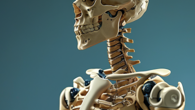
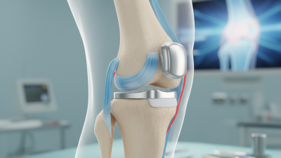
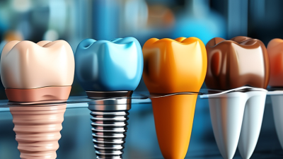
What Are Freedom of Movement Implants and How Do They Work?

Freedom of movement implants are medical devices designed to support movement when a damaged joint or bone can no longer function normally. The phrase may describe advanced orthopedic implants, including joint replacements and motion-preserving devices. It is not always a single, universally defined product category. That distinction matters.

These implants commonly use materials such as titanium alloys, cobalt-chromium, ceramics, or medical-grade polymers. Their design aims to recreate controlled movement between body structures. A hip implant, for example, may include a rounded head, a socket, and a stem fixed inside the thighbone. Each part must work with muscles, ligaments, and surrounding tissues.

Movement is the goal.

Before surgery, specialists review X-rays, CT scans, symptoms, physical limitations, and overall health. They may also consider bone quality and the patient’s daily activities. During implantation, careful positioning helps balance stability, comfort, and range of motion. Even a small alignment difference can affect walking, bending, or lifting.

Freedom of movement implants do not guarantee normal movement. Recovery often requires supervised physiotherapy, gradual strengthening, and realistic expectations. Results can vary because healing, tissue condition, implant design, and surgical technique all influence function. Some people regain comfortable movement; others may continue experiencing stiffness or pain.

Evidence and professional guidance should shape treatment decisions. Patients should ask about expected benefits, possible complications, implant lifespan, and follow-up care. Questions are valuable. A device that sounds highly advanced may still have limitations.

The best choice is personal.

This article explains how Freedom of movement implants work, why clinicians use them, and what patients should understand before considering treatment. Information cannot replace an evaluation from a qualified orthopedic specialist.
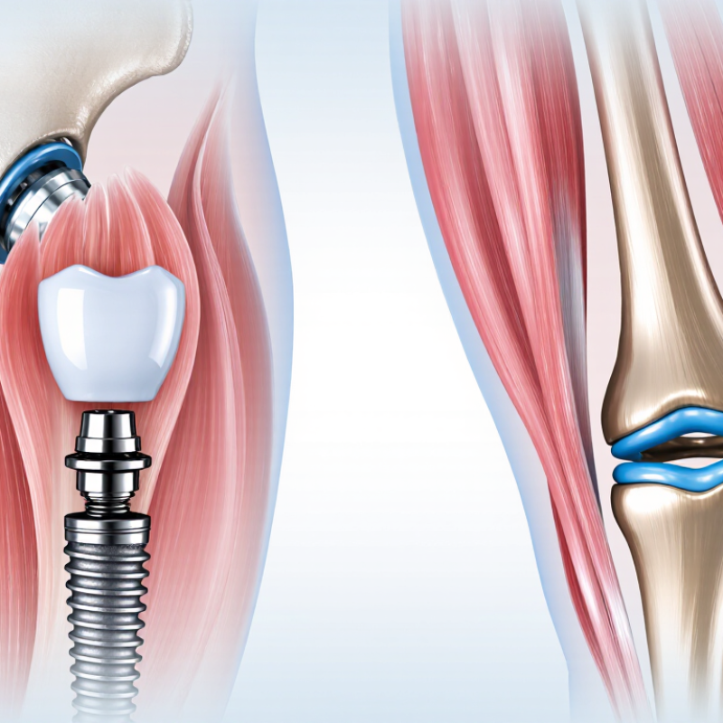
What Are Freedom of Movement Implants and How Do They Work?

Definition and Purpose of Freedom of Movement Implants

What Are Freedom of Movement Implants and How Do They Work?

Freedom of movement implants are medical devices designed to support movement instead of eliminating it. The term is not a single, universally defined implant category. It commonly describes artificial discs, mobile joint components, and motion-preserving spinal devices. Their purpose is practical: maintain controlled movement, reduce abnormal stress, and support everyday activities.

These implants use engineered surfaces that slide, rotate, or cushion under load. An artificial disc, for example, separates spinal bones while allowing limited bending. A mobile joint component can shift slightly during walking. Careful sizing matters. So does accurate placement. Surgeons assess bone quality, alignment, symptoms, and long-term expectations before recommending treatment. The implant does not create normal anatomy. It only attempts to reproduce selected mechanical functions.

The need is substantial. The World Health Organization reported that about 1.71 billion people live with musculoskeletal conditions worldwide in 2022. OECD Health at a Glance 2023 also identified hip and knee replacement as major procedures, while showing wide differences in treatment rates between countries. These figures suggest strong demand, but demand is not proof of suitability. Clinical studies still report risks, including wear, loosening, nerve irritation, and later revision surgery. Outcomes can vary with age, activity, surgical technique, and rehabilitation. The phrase “freedom of movement” can sound too optimistic. Movement may improve, but it may also remain limited. Patients should review registry data, clinical evidence, and device-specific follow-up with a qualified specialist.

What Are Freedom of Movement Implants?

Freedom of movement implants are medical devices designed to replace or support a damaged joint or spinal structure while preserving the movement that the anatomy is intended to provide. The chart shows approximate maximum movement values for healthy adult joints commonly treated with motion-restoring implants.

Approximate clinical reference values in degrees; actual movement varies by individual, diagnosis, surgical technique, rehabilitation, and implant design.

Key Components and Materials Used in These Implants

Freedom of movement implants are orthopedic devices designed to restore controlled motion in damaged joints. They usually include a bearing surface, a structural body, and a fixation system. The bearing surface allows two components to move against each other. Its shape and smoothness influence comfort, stability, and wear over time.

Material selection matters greatly. Titanium alloys often form stems, screws, or shells because they are strong and relatively light. Cobalt-chromium alloys may support highly durable joint surfaces. Ceramics can provide a very smooth bearing surface and resist scratching. Ultra-high-molecular-weight polyethylene is also used as a low-friction spacer in many joint designs. Each material has limits. Ceramics can be vulnerable to sudden impact, while polymers may gradually produce wear particles.

Some implants use porous metal surfaces to encourage bone attachment. Others rely on bone cement for immediate fixation. The choice depends on bone quality, age, activity level, anatomy, and surgical judgment. In clinical practice, even a small alignment difference can affect movement. That detail is easy to underestimate. Surgeons also examine imaging and patient history before selecting component size and placement. No implant is perfect. Movement may improve, but strength, balance, and rehabilitation still shape the result. Careful follow-up helps identify loosening, unusual wear, or restricted motion before these problems become severe.

What Are Freedom of Movement Implants and How Do They Work? - Key Components and Materials Used in These Implants

Implant Component Primary Function Common Materials How It Supports Movement Key Design or Clinical Considerations
Femoral stem Anchors the femoral side of a hip implant inside the thigh bone. Titanium alloys; cobalt-chromium alloys; stainless steel in selected designs. Provides a stable base for the femoral neck and ball while allowing the hip joint to rotate and flex. Stem geometry, offset, alignment, and fixation method influence leg length, joint stability, and biomechanics.
Femoral head Acts as the spherical moving surface of a total hip replacement. Cobalt-chromium alloy; ceramic materials such as alumina or zirconia-toughened alumina. Rotates inside the acetabular liner, enabling hip flexion, extension, abduction, adduction, and rotation. Head diameter affects jump distance, stability, range of motion, and wear behavior.
Acetabular cup Replaces the damaged hip socket and holds the bearing liner. Porous titanium or titanium alloy; cobalt-chromium alloy in some constructions. Maintains the position of the socket while permitting the femoral head to move through multiple planes. Cup inclination, version, fixation, and bone coverage affect impingement risk and stability.
Acetabular liner Creates the low-friction bearing surface against the femoral head. Highly cross-linked ultra-high-molecular-weight polyethylene; ceramic in selected ceramic-on-ceramic systems. Allows smooth articulation and helps distribute contact forces during walking and other activities. Thickness, conformity, wear resistance, and resistance to edge loading are important factors.
Knee femoral component Resurfaces the lower end of the femur in total knee replacement. Cobalt-chromium alloy; oxidized zirconium in selected designs. Provides smooth curved surfaces for flexion and extension and guides knee motion. Femoral radius, component rotation, and ligament balancing influence stability and functional movement.
Knee tibial tray Provides the structural platform on the upper surface of the tibia. Titanium alloy; cobalt-chromium alloy in some designs. Supports the polyethylene insert and transfers load from the femur to the tibia. Alignment, fixation, tibial slope, and rotational positioning affect kinematics and load distribution.
Knee polyethylene insert Separates the metal femoral and tibial components and provides the main bearing surface. Ultra-high-molecular-weight polyethylene, commonly highly cross-linked or otherwise wear-optimized. Permits controlled sliding, rolling, and limited rotation while reducing metal-to-metal contact. Insert thickness, conformity, constraint level, and polyethylene wear affect motion and longevity.
Patellar component Resurfaces the underside of the kneecap when patellar resurfacing is performed. Ultra-high-molecular-weight polyethylene. Glides against the femoral component during knee bending and straightening. Component position and patellar tracking are important for smooth movement and anterior knee comfort.
Fixation interface Secures the implant to bone either with cement or through biological bone ingrowth. Bone cement based on polymethyl methacrylate; porous titanium or tantalum for cementless fixation. Maintains component alignment so the joint can move under repeated loading without excessive micromotion. Bone quality, implant geometry, surface porosity, and surgical technique influence fixation security.
Modular connection Joins interchangeable parts such as a hip stem and femoral head or a knee tray and insert. Titanium alloys, cobalt-chromium alloys, and compatible polymer components. Allows surgeons to adjust dimensions, offset, stability, and soft-tissue tension during reconstruction. Connection strength, taper fit, corrosion resistance, and compatibility between components must be carefully controlled.

Note: “Freedom of movement implants” is a descriptive term rather than a single standardized implant category. Actual range of motion depends on implant geometry, fixation, soft-tissue condition, bone anatomy, surgical positioning, rehabilitation, and individual patient factors.

How the Implant Restores or Supports Natural Movement

Freedom of movement implants are designed to restore useful motion when a damaged joint can no longer move smoothly. Osteoarthritis affected about 528 million people worldwide in 2019, according to the World Health Organization. For many patients, joint replacement can reduce painful bone-on-bone contact and support daily activities such as walking, climbing stairs, or standing from a chair.

The implant usually combines smooth, shaped surfaces with a durable spacer. During movement, these parts guide the joint through a controlled arc. Muscles and ligaments still matter. They provide stability, power, and body awareness after surgery. The implant does not create natural movement by itself. It gives the surrounding tissues a better mechanical environment.

Small details matter.

Surgeons select component size, alignment, and positioning from imaging and physical examination. Rehabilitation then rebuilds strength and coordination through repeated, measured movements.

Data from the National Joint Registry’s 21st Annual Report, covering more than three million recorded joint procedures, shows why long-term monitoring remains important. Implant survival can be strong, but results vary with age, activity, bone quality, surgical technique, and rehabilitation. That uncertainty deserves honesty. A patient may regain comfortable motion without recovering every movement experienced in youth. The real goal is often practical independence, not perfect anatomy.

Surgical Placement, Adjustment, and Recovery Process

Freedom of movement implants are medical devices designed to support mobility while preserving controlled motion. Their exact design depends on the joint, injury, and patient’s anatomy. A qualified surgeon reviews imaging, movement limits, bone quality, and daily activities before recommending one. The implant should fit the person, not just the scan.

During surgery, the patient receives anesthesia and the area is prepared under sterile conditions. The surgeon removes damaged tissue when necessary, then places the implant components with careful alignment. Trial parts may be used first. This allows the team to assess stability, range of motion, and soft-tissue tension before final placement. Small adjustments can matter.

Afterward, swelling and stiffness are common. Recovery usually begins with guided exercises and short, supported movements. A therapist may teach safe walking, transfers, and home exercises. Some adjustable implants can be modified by a clinician during follow-up visits. Patients should never attempt to change an implant themselves.

Progress is not always smooth. One week may feel stronger than the next. Pain, warmth, drainage, fever, or sudden loss of movement needs prompt medical attention. Follow-up visits help assess healing and implant position. Recovery also depends on sleep, nutrition, existing conditions, and honest communication with the care team. Healing takes time.

Benefits, Risks, and Long-Term Care Considerations

Freedom of movement implants are orthopedic devices designed to restore or preserve controlled joint motion. They may replace damaged surfaces or support a motion segment, depending on the body area. Their polished bearing surfaces and shaped components guide movement while transferring load through bone. Proper alignment matters greatly. A few millimeters can change comfort, stability, and walking mechanics.

Potential benefits include reduced pain, improved range of motion, and greater independence during daily activities. The 2024 National Joint Registry recorded more than three million joint replacement procedures in England and Wales. Its data show revision risk varies by joint, implant design, fixation method, age, and sex. No device offers a guaranteed lifetime result. That point is often underexplained.

Risks include infection, loosening, fracture, dislocation, wear debris, nerve injury, and stiffness. Some patients gain motion but still experience weakness or persistent pain. Evidence is not perfectly clean. Long-term follow-up can reveal problems that short clinical studies miss. The FDA emphasizes post-market surveillance because real-world performance may differ from trial results.

Care usually includes gradual strengthening, weight management, fall prevention, and scheduled clinical reviews. Patients should report increasing pain, swelling, warmth, instability, or reduced movement. Imaging may detect migration or wear before symptoms become severe. Avoiding every high-impact activity is not always necessary, but activity should match the surgeon’s advice. Follow-up should continue even when movement feels normal.

volume-unmute-yellow-icon volume-unmute-blue-icon