Companies




Browse products by medical specialities area or therapies

Arthroplasty
Product by Therapies
Product by Type
Sports Medicine
Product by Therapies
Knee Portfolio - ACL /PCL Reconstruction
Knee Portfolio - Meniscus Repair
Shoulder Portfolio - Bankart/Slap Repair
Shoulder Portfolio - Rotator Cuff Repair
Product by Type


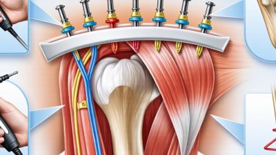
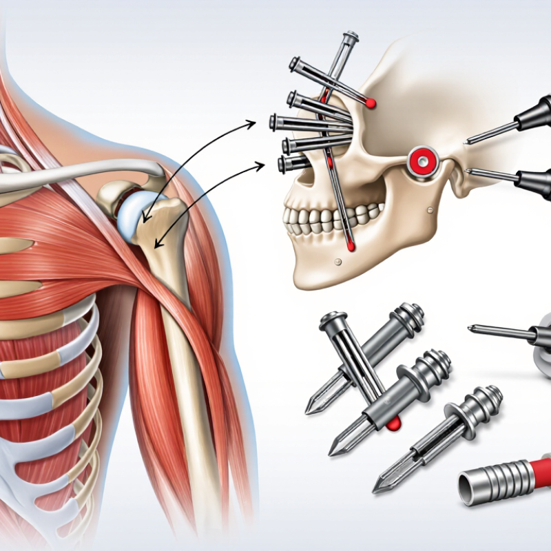
Choosing Shoulder Bankart repair systems is not a decision based on hardware alone. It involves anatomy, injury pattern, surgical technique, and the patient’s recovery goals. A well-designed system can help surgeons address anterior shoulder instability with compatible anchors, instruments, and procedural options.
Precision matters. During repair, the surgeon may work through small portals while viewing the glenoid rim on a monitor. Reliable instruments can support controlled anchor placement and secure soft-tissue fixation. Some systems also offer ergonomic handles, clear depth guidance, and configurations suited to different surgical preferences. These details may reduce unnecessary handling during a demanding procedure.
But no system guarantees a successful outcome. That point deserves attention. Results depend on accurate diagnosis, appropriate patient selection, tissue quality, surgeon experience, and structured rehabilitation. Current clinical reasoning should guide the choice, not marketing language. Surgeons should review published evidence, device specifications, material properties, and training requirements before adopting a platform.
A practical evaluation also considers workflow. Does the system provide consistent instrumentation? Can the team prepare it efficiently? Are the components compatible with the intended technique? These questions influence operating-room confidence and procedural reliability. They also reveal limitations that promotional descriptions may overlook.
For patients, the discussion should remain clear and realistic. Shoulder Bankart repair systems may support a careful repair, but they do not replace clinical judgment. Their value lies in helping qualified professionals perform a planned procedure with control and consistency. The best choice is often the one that fits the patient, the surgeon, and the evidence—not simply the newest option.
Why Choose Shoulder Bankart Repair Systems?
A shoulder Bankart lesion is a tear of the labrum along the front-lower socket. It often follows a dislocation, fall, or contact-sport injury. The torn tissue can reduce shoulder stability. Some patients describe a sudden slipping sensation. Others notice pain, clicking, weakness, or fear during overhead movement. Symptoms vary widely. That makes diagnosis more careful than it first appears.
A clinician usually reviews the injury, movement limits, and previous dislocations. Physical tests may reproduce apprehension or instability. X-rays can help exclude fractures and alignment problems. MRI or MR arthrography may show labral damage and related soft-tissue injuries. No single test explains every shoulder complaint. In practice, symptoms and images sometimes disagree, and that deserves honest discussion. Treatment decisions should consider age, activity, bone loss, tissue quality, and recurrence risk. Shoulder Bankart repair systems can help secure the repaired labrum, but the device is only one part of the procedure. Surgical experience and rehabilitation also influence recovery.
Tips: Keep the shoulder protected after injury. Avoid forcing painful positions. Record when instability occurs and what movement triggered it. Seek assessment after a first-time dislocation or repeated slipping. A second opinion can be useful when imaging and symptoms do not match. Ask how the repair system fits your anatomy, what complications are possible, and how long rehabilitation may take. Recovery is not always linear. Expect adjustments.
| Clinical Dimension | Feature | Evidence-Based Information | Clinical Significance | Relevance to a Bankart Repair System |
|---|---|---|---|---|
| Lesion Definition | Typical anatomical injury | A Bankart lesion is a detachment of the anteroinferior glenoid labrum from the rim of the glenoid cavity. The injury may also involve the adjacent capsule and inferior glenohumeral ligament complex. | Loss of the normal soft-tissue stabilizing mechanism can contribute to recurrent anterior shoulder instability. | A suitable system should support secure fixation of the labrum and capsule while allowing accurate placement along the anterior and inferior glenoid rim. |
| Common Causes | Traumatic anterior shoulder dislocation | The lesion most commonly occurs when the humeral head dislocates anteriorly, particularly during forced abduction and external rotation. | A traumatic dislocation may stretch or detach the labrum and may be associated with capsular laxity, glenoid bone loss, or a Hill-Sachs lesion. | The repair construct should accommodate soft-tissue tensioning and allow stable fixation in cases with different tear locations and tissue quality. |
| Risk Factors | Age, activity, and recurrence | Younger patients and individuals participating in contact, collision, or overhead sports generally have a higher risk of recurrent instability after an initial traumatic event. | Recurrent episodes can enlarge the lesion, increase capsular attenuation, and contribute to progressive bone loss. | A versatile system can facilitate reproducible fixation across multiple anchor positions and support treatment planning based on patient activity and tissue condition. |
| Typical Symptoms | Pain and apprehension | Common symptoms include anterior shoulder pain, a feeling of instability, apprehension when the arm is elevated and externally rotated, and a history of slipping or dislocation. | Symptoms may persist even when strength and range of motion appear relatively normal between instability episodes. | Reliable fixation may help restore the labral bumper and capsuloligamentous tension, which are important components of anterior stability. |
| Physical Examination | Instability testing | The apprehension, relocation, and release tests may reproduce symptoms. Assessment should also include generalized laxity, range of motion, rotator cuff strength, and neurovascular status. | Examination findings help distinguish instability from rotator cuff disease, acromioclavicular pathology, or cervical-related symptoms. | Surgical planning should match the lesion pattern identified through examination and imaging rather than relying on a single standard configuration. |
| Plain Radiography | Initial imaging | Anteroposterior, axillary, and specialized shoulder views can identify dislocation, fracture, degenerative change, and some osseous defects. | Radiographs are useful for detecting associated bony injury but may not fully demonstrate the labral tear. | Findings such as glenoid rim fracture or humeral head impaction may influence whether soft-tissue repair alone is appropriate. |
| Magnetic Resonance Imaging | Labral and soft-tissue assessment | MRI can evaluate the labrum, capsule, rotator cuff, cartilage, and associated bone lesions. MR arthrography may improve visualization of intra-articular labral pathology. | Imaging helps define tear location, extension, tissue quality, and associated injuries before surgery. | A repair system should provide options for different tear extents and permit controlled reattachment of the labrum and capsule under arthroscopic visualization. |
| Computed Tomography | Bone-loss evaluation | CT, particularly three-dimensional reconstruction, is useful when glenoid bone loss, a bony Bankart lesion, or a clinically important Hill-Sachs lesion is suspected. | The amount and location of bone loss can affect procedure selection and may make isolated soft-tissue repair less suitable. | A Bankart repair system is most appropriate when the principal pathology is repairable soft-tissue detachment and the bony anatomy is suitable for the planned procedure. |
| Repair Objective | Restoration of anatomy | The goals are to reattach the detached labrum, restore the anterior capsulolabral complex, and improve resistance to anterior translation of the humeral head. | Anatomical restoration is intended to reduce recurrent instability while preserving functional shoulder motion. | Important design considerations include secure fixation, predictable deployment, appropriate anchor size, and compatibility with the planned number and location of fixation points. |
| System Selection | Key technical considerations | Relevant factors include anchor fixation strength, insertion control, suture management, tissue handling, visibility, and compatibility with arthroscopic instruments. | The selected system should support the surgeon’s technique and the patient’s specific anatomy rather than impose a single repair strategy. | Choosing a well-matched system may improve procedural consistency, facilitate accurate capsulolabral repair, and reduce avoidable technical difficulty. |
| Postoperative Considerations | Rehabilitation and follow-up | Rehabilitation commonly involves a period of protection, progressive range-of-motion recovery, strengthening, and gradual return to sport or demanding activity. | Recovery timelines vary according to the repair, associated injuries, healing response, occupation, and sporting demands. | A mechanically secure repair must be combined with appropriate rehabilitation; implant selection alone cannot eliminate the risk of recurrence or stiffness. |
| Clinical decisions should be based on a complete history, physical examination, appropriate imaging, patient activity level, bone and soft-tissue condition, and the treating surgeon’s judgment. This table is for general educational use and does not replace individualized medical advice. | ||||